Peripheral Vascular Disease Doppler Ultrasound
Peripheral vascular disease doppler ultrasound. The history of Doppler ultrasound in peripheral vascular diagnosis is considered in terms of basic developments clinical applications and impact on medical practice. Despite the advances in computed tomography CT and magnetic resonance MR angiographic techniques vascular ultrasound still represents the initial test of choice based on its noninvasive nature accuracy validated values and wide availability. Peripheral vascular disease can affect all types of blood vessels.
Introduction Doppler ultrasound scan is a highly useful non-invasive and reasonable tool for determination of CIMT peripheral arterial diseases renal artery stenosis due to atherosclerosis caused by diabetes mellitus hyperlipidemia and smoking 1 2. While there are several types of peripheral ultrasounds each one uses ultrasonic sound waves as a means of detecting blood flow within the arteries and veins. If playback doesnt begin shortly try restarting your device.
Doppler ultrasonography of the lower extremity arteries is a valuable technique although it is less frequently indicated for peripheral arterial disease than for deep vein thrombosis or varicose veins. Peripheral vascular doppler ultrasound is a non-invasive cardiovascular testing procedure that is typically recommended for patients who are experiencing symptoms of peripheral artery disease PAD. The sonographic examination of patients with peripheral vascular disease will in general complement the use of other physiologic tests such as pressure measurements plethysmographic recordings and contin-uous wave Doppler.
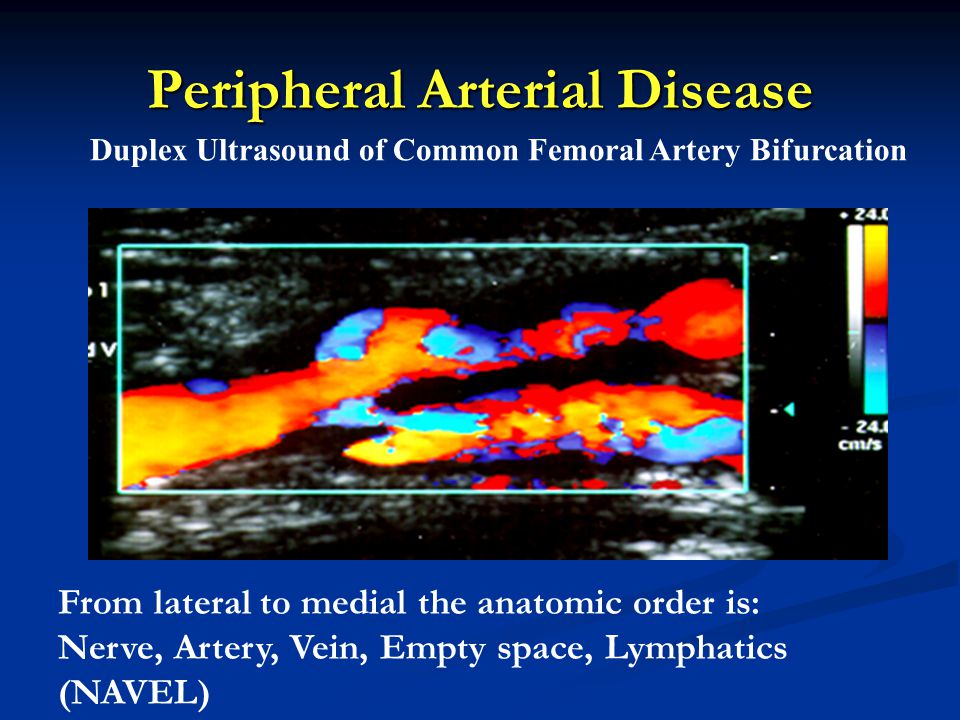
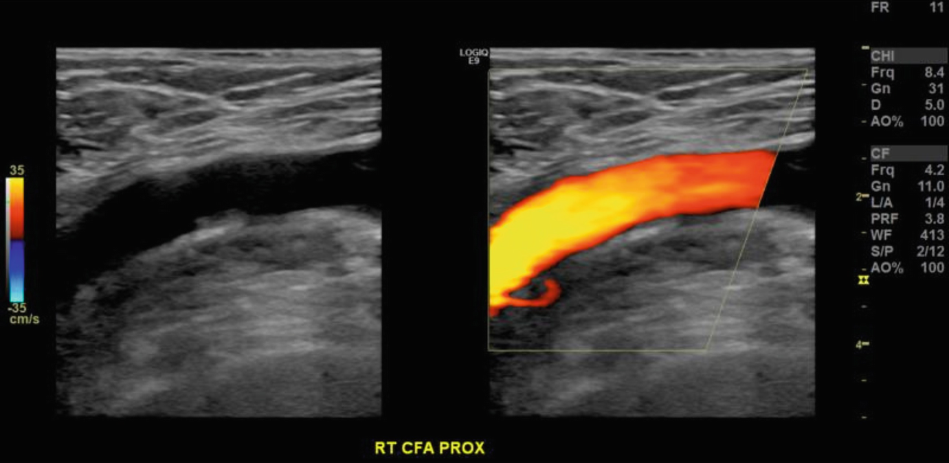
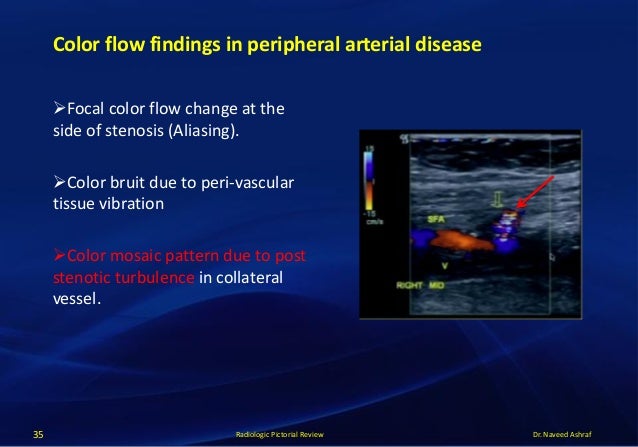
The ankle brachial pressure index is a relatively simple method for quantifying the severity of arterial occlusion in the leg. The most common symptom is pain which becomes worse as the circulation more limited. Performing noninvasive evaluation of the peripheral arteries using color and Doppler waveform analysis ultrasound.
Doppler Evaluation of Peripheral Arterial Disease. Consult with a cardiologist to see if this test is needed for you. This disease more often affects the blood vessels in the legs.
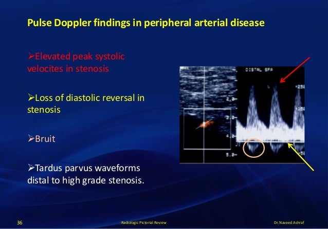
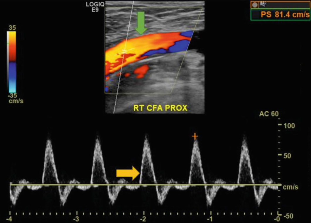
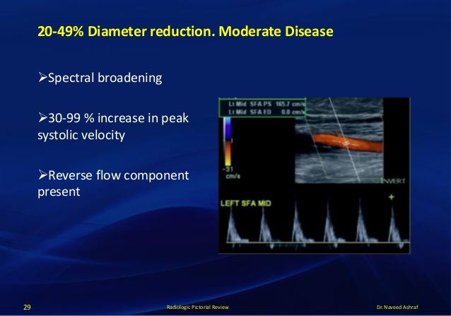
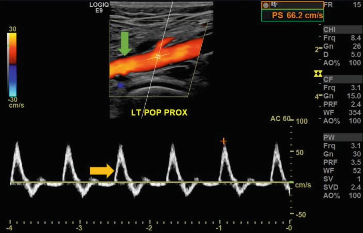
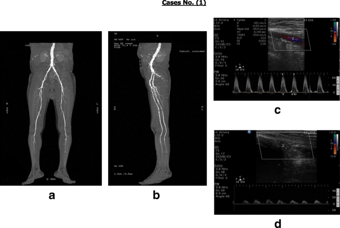
A blood pressure cuff is inflated around the lower calf muscle above the ankle joint and a doppler ultrasound probe placed over the dorsalis pedis artery and the posterior tibial artery. A Doppler Ultrasound is a test that estimates the blood flow through your body. Spectral Doppler waveform assessment has been the principal diagnostic tool for the noninvasive diagnosis of peripheral arterial and venous disease since the 1980s.
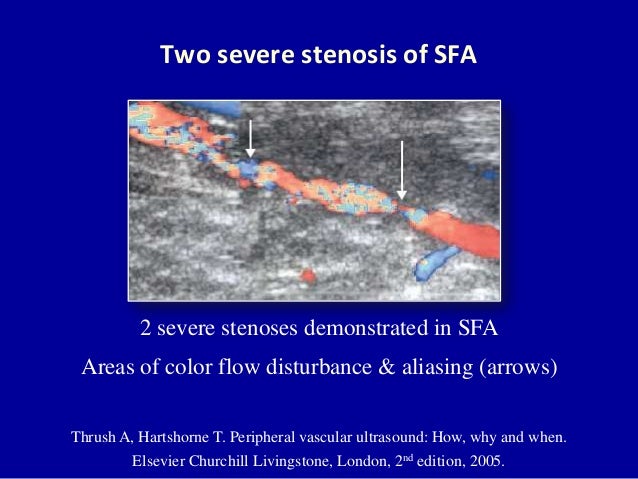
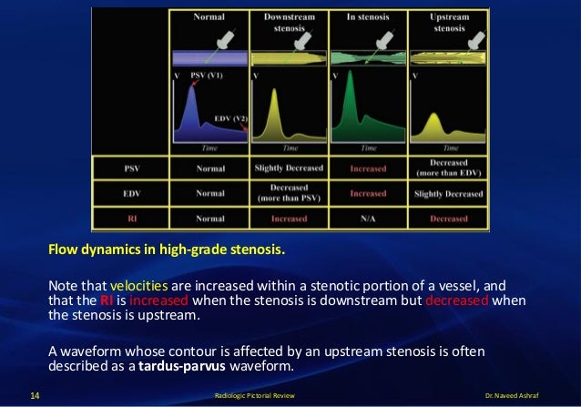
3637 The absence of uniformly accepted standardized descriptors for arterial and venous Doppler waveforms has been a long-standing point of controversy 38 and the main impetus for the development of this consensus. Ultrasonography can diagnose stenosis through the direct visualization of plaques and.
PAD Peripheral Arterial Diseases Doppler Ultrasonography T2DM Type 2Diabetes Mellitus 1.
While there are several types of peripheral ultrasounds each one uses ultrasonic sound waves as a means of detecting blood flow within the arteries and veins. Introduction Doppler ultrasound scan is a highly useful non-invasive and reasonable tool for determination of CIMT peripheral arterial diseases renal artery stenosis due to atherosclerosis caused by diabetes mellitus hyperlipidemia and smoking 1 2. While there are several types of peripheral ultrasounds each one uses ultrasonic sound waves as a means of detecting blood flow within the arteries and veins. Despite the advances in computed tomography CT and magnetic resonance MR angiographic techniques vascular ultrasound still represents the initial test of choice based on its noninvasive nature accuracy validated values and wide availability. Peripheral vascular disease can affect all types of blood vessels. Blood flow is restricted to the tissue because of spasm or narrowing of the vessel. This disease more often affects the blood vessels in the legs. The history of Doppler ultrasound in peripheral vascular diagnosis is considered in terms of basic developments clinical applications and impact on medical practice. A blood pressure cuff is inflated around the lower calf muscle above the ankle joint and a doppler ultrasound probe placed over the dorsalis pedis artery and the posterior tibial artery.
Blood flow is restricted to the tissue because of spasm or narrowing of the vessel. Many early developments occurred at Osaka University in Japan and the University of Washington in the United States. If playback doesnt begin shortly try restarting your device. Doppler ultrasonography of the lower extremity arteries is a valuable technique although it is less frequently indicated for peripheral arterial disease than for deep vein thrombosis or varicose veins. Vascular ultrasound provides a valuable tool in the assessment of various peripheral vascular beds. Performing noninvasive evaluation of the peripheral arteries using color and Doppler waveform analysis ultrasound. Despite the advances in computed tomography CT and magnetic resonance MR angiographic techniques vascular ultrasound still represents the initial test of choice based on its noninvasive nature accuracy validated values and wide availability.



















Post a Comment for "Peripheral Vascular Disease Doppler Ultrasound"